|
|
| (21 intermediate revisions not shown) |
| Line 1: |
Line 1: |
| - | __TOC__
| |
| - | === Goal ===
| |
| - |
| |
| | <html> | | <html> |
| - | <h4>To understand how the mind, behavior and brain are interrelated by linking key cognitive & emotional functions to neural activities at multiple levels of brain systems. <h4>
| |
| - | <img src="http://bcs.snu.ac.kr/mediawiki/uploads/e/e0/Goal.png" alt="Goal" width="700" height="500"></p>
| |
| - | </html>
| |
| - | <!--
| |
| - | <p align=center><img src="/preview/img/mind.png" alt="Research" /></p>
| |
| - | -->
| |
| - | === Plans ===
| |
| - | ====The Strategy of Linking Brain and Mind (SLBM)====
| |
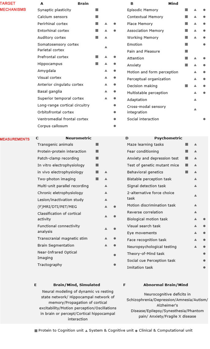
| - | First, we have identified a set of comprehensive but focused set of cognitive functions and targeted
| |
| - | brain mechanisms with an aim to achieve breadth while insuring that our studies fall squarely within the
| |
| - | areas of expertise of our associated faculty (Fig 1A, 1B). Second, to address the 'linking' questions
| |
| - | around those key cognitive and brain mechanisms, we intend to integrate cutting-edge ‘neurometric'
| |
| - | techniques to probe neural activities/structures at multiple levels spanning the depth of genetic, molecular,
| |
| - | synaptic, neuronal, local/global network-level measurements (Fig 1C). These brain measurements will be
| |
| - | paralleled by state-of-the-art 'psychometric' tools to capture mind in action, with behavioral tasks
| |
| - | spanning the breadth of behavioral genetics, rodent memory tasks, visual perception tasks, eye movements,
| |
| - | reverse correlation and social perception tasks (Fig 1D). Third, based on the neurometric and
| |
| - | psychometric data, we will build tight linkages between brain activity/structure and mind. In doing so,
| |
| - | computational approaches will help us to build comprehensive neural models constrained by data from
| |
| - | empirical studies (Fig 1E). Computational models of complex brain systems allow us to put to test, via
| |
| - | simulation, predictions that cannot be tested in empirical situations and to guide empirical studies by
| |
| - | generating testable predictions about behaviors in the domains of both brain and mind. In addition, we
| |
| - | will study brains and minds in abnormal states by specifying the roles of neural circuits underlying
| |
| - | cognitive deficits in patients with psychiatric disorders and in individuals with remarkable cognitive
| |
| - | abilities that fall outside the range of normal (Fig 1F). Our multi-level approach, transpiring through
| |
| - | 'gene', 'protein', 'neuron', 'system', 'behavior', 'abnormality' and 'computation', can lead to
| |
| - | fundamental cure for mental diseases in the future.
| |
| | | | |
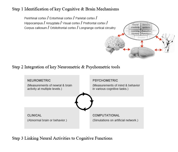
| - | ==== <font color="#000">step 1</font> Identification of key ''Cognitive'' & ''Brain mechanisms'' ====
| + | <table cellspacing=0 cellpadding=0 width="900" border="0"> |
| - | <html>
| + | <tr><td colspan="3"><img alt="" src="/mediawiki/uploads/8/8d/Img_blank1.jpg" width="38" height="38"></td></tr> |
| - | <table width=770 border="0" cellpadding="0" cellspacing="0"> | + | <tr><td width="131"></td> |
| - | <tr><td align=center> | + | <td width="769"> |
| - | Perirhinal cortex / Entorhinal cortex / Parietal cortex /<br>
| + | |
| - | Hippocampus / Amygdala / Visual cortex / Prefrontal<br>
| + | |
| - | cortex / Corpus callosum / Orbitofrontal cortex / Longrange<br>
| + | |
| - | cortical circuitry
| + | |
| - | </td>
| + | |
| - | <td align=center valign=middle><br>
| + | |
| - | <img src=/preview/img/strategic.gif alt="Strategic Plans For Research" border=0>
| + | |
| - | </td>
| + | |
| - | </tr> | + | |
| - | </table>
| + | |
| - | </html>
| + | |
| | | | |
| - | ==== <font color="#000">step 2</font> Integration of key Neurometric & Psychometric tools ==== | + | <table width="100%" cellspacing=0 cellpadding=0 border=0> |
| - | <html> | + | <tr><td colspan=2><img src="/mediawiki/uploads/c/c6/02_title.jpg"></td></tr> |
| - | <p align="center"><img src="http://bcs.snu.ac.kr/preview/img/step2.gif" alt="step 2 integration"></p> | + | <tr><td colspan=2><img src="/mediawiki/uploads/5/5c/02_img01.jpg" alt=""></td></tr> |
| - | </html> | + | |
| | + | <tr><td><span style="text-transform:uppercase;color:#000000;font-size:17px;font-family:arial;font-weight:bold">The strategy of linking brain and mind (SJRM)</span></td></tr> |
| | + | <tr><td height=20> </td><td rowspan=20 width=35><img src="/mediawiki/uploads/5/5a/Blank2.gif"></td></tr> |
| | + | <tr><td span style="font-family:arial;font-size:12px;color:#414141;line-height:16px;text-align:justify"><b>First,</b> we have identified a set of comprehensive but focused set of cognitive functions and targeted brain mechanisms with an aim to achieve breadth while insuring that our studies fall squarely within the areas of expertise of our associated faculty. |
| | + | <p><br /></p><b>Second,</b> to address the 'linking' questions around those key cognitive and brain mechanisms, we intend to integrate cutting-edge 'neurometric' techniques to probe neural activities/structures at multiple levels spanning the depth of genetic, molecular, synaptic, neuronal/global network-level measurements. These brain measurements will be paralleled by state-of-the-art 'psychometric' tools to capture in mind in action, with behavioral tasks spanning the breadth of behavioral genetics, rodent memory tasks, visual perception tasks, eye movements, reverse correlation and social perception tasks. Third, based on the neurometric and psychometric data, we will build tight linkages between brain activity/structure and mind. In doing so, computational approaches will help us to build comprehensive neural models constrained by data from empirical studies. Computational models of complex brain systems allow us to put to test, via simulation, predictions that cannot be tested in empirical situations and to guide empirical studies by generating testable predictions about behaviors in the domains of both brain and mind. |
| | | | |
| - | ==== <font color="#000">step 3</font> Linking Neural Activities to Cognitive Functions ====
| + | <p><br /></p>In addition, we will study brains and minds in abnormal states by specifying the roles of neural circuits underlying cognitive deficits in patients with psychiatric disorders and in individuals with remarkable cognitive abilities that fall outside the range of normal.<br /> |
| - | <br/> | + | Our multi-level approach, transpiring through 'gene', 'protein', 'neuron', 'system', 'behavior', 'abnormality' and 'computation', can lead to fundamental cure for mental diseases in the future.</td></tr> |
| - | <br/> | + | |
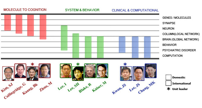
| - | === Research Units ===
| + | |
| - | ==== Three Core Research Units ====
| + | |
| - | <html><p align="center"><img src="/preview/img/organization.png" alt="Organization" /></p></html>
| + | |
| - | <br/> | + | |
| - | <br/> | + | |
| - | <br/> | + | |
| | | | |
| - | === Research overview ===
| + | <tr><td height=10> </td></tr> |
| - | <html>
| + | <tr><td><img src="/mediawiki/uploads/5/52/02_img02.jpg" alt=""></td></tr> |
| - | <!--Research-->
| + | |
| - | <table width="780" border="0" cellpadding="0" cellspacing="0">
| + | |
| - | <tr> | + | |
| - | <td align=left><font color=000000><B>I. Introduction and overview of research plans</B></font><br>
| + | |
| - | Our research aim is to use biological, behavioral and computational methods to understand how the
| + | |
| - | mind, brain and behavior are interrelated. Studying humans and animals, our research will link key
| + | |
| - | cognitive and emotional functions to neural activities at multiple levels of brain systems. To establish
| + | |
| - | these linkages, we set forth a research strategy, named 'SLBM (Strategy of Linking Brain and Mind)', the
| + | |
| - | major steps and components of which are summarized in the following three sections.
| + | |
| - | <br><br>
| + | |
| - | <table width=760 border="0" cellpadding="0" cellspacing="0">
| + | |
| - | <tr><td align=center valign=middle rowspan=2 class="research1" width=110><b>Target<br> mechanisms</b></td>
| + | |
| - | <td align=left>
| + | |
| - | <table width=300 border="0" cellpadding="0" cellspacing="0">
| + | |
| - | <tr><td align=center class=research1><b>A </b></td>
| + | |
| - | <td align=center class=research1><b>Brain</b></td></tr>
| + | |
| - | </table>
| + | |
| - | </td>
| + | |
| - | <td align=left>
| + | |
| - | <table width=300 border="0" cellpadding="0" cellspacing="0">
| + | |
| - | <tr><td align=center class=research1><b>B </b></td>
| + | |
| - | <td align=center class=research1><b>Mind</b></td></tr>
| + | |
| - | </table>
| + | |
| - | </td>
| + | |
| - | </tr>
| + | |
| - | <tr><td align=center>
| + | |
| - | <table width=300 border="0" cellpadding="2" cellspacing="1" bgcolor=dbdbdb>
| + | |
| - | <tr><td align=left bgcolor=ffffff width=210>Synaptic plasticity</td>
| + | |
| - | <td align=center bgcolor=ffffff width=30><img src=/preview/img/square.png border=0></td>
| + | |
| - | <td align=center bgcolor=ffffff width=30> </td>
| + | |
| - | <td align=center bgcolor=ffffff width=30> </td></tr>
| + | |
| - | <tr><td align=left bgcolor=ffffff>Calcium sensors</td>
| + | |
| - | <td align=center bgcolor=ffffff><img src=/preview/img/square.png border=0></td>
| + | |
| - | <td align=center bgcolor=ffffff></td>
| + | |
| - | <td align=center bgcolor=ffffff></td></tr>
| + | |
| - | <tr><td align=left bgcolor=ffffff>Perirhinal cortex</td>
| + | |
| - | <td align=center bgcolor=ffffff><img src=/preview/img/square.png border=0></td>
| + | |
| - | <td align=center bgcolor=ffffff><img src=/preview/img/triangle.png border=0></td>
| + | |
| - | <td align=center bgcolor=ffffff><img src=/preview/img/circle.png border=0></td></tr>
| + | |
| - | <tr><td align=left bgcolor=ffffff>Entorhinal cortex</td>
| + | |
| - | <td align=center bgcolor=ffffff><img src=/preview/img/square.png border=0></td>
| + | |
| - | <td align=center bgcolor=ffffff><img src=/preview/img/triangle.png border=0></td>
| + | |
| - | <td align=center bgcolor=ffffff><img src=/preview/img/circle.png border=0></td></tr>
| + | |
| - | <tr><td align=left bgcolor=ffffff>Auditory cortex</td>
| + | |
| - | <td align=center bgcolor=ffffff><img src=/preview/img/square.png border=0></td>
| + | |
| - | <td align=center bgcolor=ffffff><img src=/preview/img/triangle.png border=0></td>
| + | |
| - | <td align=center bgcolor=ffffff></td></tr>
| + | |
| - | <tr><td align=left bgcolor=ffffff>Somatosensory cortex
| + | |
| - | Parietal cortex</td>
| + | |
| - | <td align=center bgcolor=ffffff></td>
| + | |
| - | <td align=center bgcolor=ffffff><img src=/preview/img/triangle.png border=0></td>
| + | |
| - | <td align=center bgcolor=ffffff></td></tr>
| + | |
| - | <tr><td align=left bgcolor=ffffff>Prefrontal cortex</td>
| + | |
| - | <td align=center bgcolor=ffffff><img src=/preview/img/square.png border=0></td>
| + | |
| - | <td align=center bgcolor=ffffff><img src=/preview/img/triangle.png border=0></td>
| + | |
| - | <td align=center bgcolor=ffffff><img src=/preview/img/circle.png border=0></td></tr>
| + | |
| - | <tr><td align=left bgcolor=ffffff>Hippocampus</td>
| + | |
| - | <td align=center bgcolor=ffffff><img src=/preview/img/square.png border=0></td>
| + | |
| - | <td align=center bgcolor=ffffff><img src=/preview/img/triangle.png border=0></td>
| + | |
| - | <td align=center bgcolor=ffffff><img src=/preview/img/circle.png border=0></td></tr>
| + | |
| - | <tr><td align=left bgcolor=ffffff>Amygdala</td>
| + | |
| - | <td align=center bgcolor=ffffff></td>
| + | |
| - | <td align=center bgcolor=ffffff><img src=/preview/img/triangle.png border=0></td>
| + | |
| - | <td align=center bgcolor=ffffff><img src=/preview/img/circle.png border=0></td></tr>
| + | |
| - | <tr><td align=left bgcolor=ffffff>Visual cortex</td>
| + | |
| - | <td align=center bgcolor=ffffff></td>
| + | |
| - | <td align=center bgcolor=ffffff><img src=/preview/img/triangle.png border=0></td>
| + | |
| - | <td align=center bgcolor=ffffff><img src=/preview/img/circle.png border=0></td></tr>
| + | |
| - | <tr><td align=left bgcolor=ffffff>Anterior cingulatu cortex</td>
| + | |
| - | <td align=center bgcolor=ffffff></td>
| + | |
| - | <td align=center bgcolor=ffffff><img src=/preview/img/triangle.png border=0></td>
| + | |
| - | <td align=center bgcolor=ffffff><img src=/preview/img/circle.png border=0></td></tr>
| + | |
| - | <tr><td align=left bgcolor=ffffff>Basal ganglia</td>
| + | |
| - | <td align=center bgcolor=ffffff></td>
| + | |
| - | <td align=center bgcolor=ffffff><img src=/preview/img/triangle.png border=0></td>
| + | |
| - | <td align=center bgcolor=ffffff><img src=/preview/img/circle.png border=0></td></tr>
| + | |
| - | <tr><td align=left bgcolor=ffffff>Superior temporal cortex</td>
| + | |
| - | <td align=center bgcolor=ffffff></td>
| + | |
| - | <td align=center bgcolor=ffffff><img src=/preview/img/triangle.png border=0></td>
| + | |
| - | <td align=center bgcolor=ffffff><img src=/preview/img/circle.png border=0></td></tr>
| + | |
| - | <tr><td align=left bgcolor=ffffff>Long-range cortical circuitry</td>
| + | |
| - | <td align=center bgcolor=ffffff></td>
| + | |
| - | <td align=center bgcolor=ffffff></td>
| + | |
| - | <td align=center bgcolor=ffffff><img src=/preview/img/circle.png border=0></td></tr>
| + | |
| - | <tr><td align=left bgcolor=ffffff>Orbitofrontal cortex</td>
| + | |
| - | <td align=center bgcolor=ffffff></td>
| + | |
| - | <td align=center bgcolor=ffffff></td>
| + | |
| - | <td align=center bgcolor=ffffff><img src=/preview/img/circle.png border=0></td></tr>
| + | |
| - | <tr><td align=left bgcolor=ffffff>Ventromedial frontal cortex</td>
| + | |
| - | <td align=center bgcolor=ffffff></td>
| + | |
| - | <td align=center bgcolor=ffffff></td>
| + | |
| - | <td align=center bgcolor=ffffff><img src=/preview/img/circle.png border=0></td></tr>
| + | |
| - | <tr><td align=left bgcolor=ffffff>Corpus callosum</td>
| + | |
| - | <td align=center bgcolor=ffffff></td>
| + | |
| - | <td align=center bgcolor=ffffff></td>
| + | |
| - | <td align=center bgcolor=ffffff><img src=/preview/img/circle.png border=0></td></tr>
| + | |
| - | </table></td>
| + | |
| - | <td align=center valign=top>
| + | |
| - | <table width=300 border="0" cellpadding="2" cellspacing="1" bgcolor=dbdbdb>
| + | |
| - | <tr><td align=left bgcolor=ffffff width=210>Episodic Memory</td>
| + | |
| - | <td align=center bgcolor=ffffff width=30><img src=/preview/img/square.png border=0></td>
| + | |
| - | <td align=center bgcolor=ffffff width=30><img src=/preview/img/triangle.png border=0></td>
| + | |
| - | <td align=center bgcolor=ffffff width=30><img src=/preview/img/circle.png border=0></td></tr>
| + | |
| - | <tr><td align=left bgcolor=ffffff>Contextual Memory</td>
| + | |
| - | <td align=center bgcolor=ffffff><img src=/preview/img/square.png border=0></td>
| + | |
| - | <td align=center bgcolor=ffffff><img src=/preview/img/triangle.png border=0></td>
| + | |
| - | <td align=center bgcolor=ffffff><img src=/preview/img/circle.png border=0></td></tr>
| + | |
| - | <tr><td align=left bgcolor=ffffff>Place Memory</td>
| + | |
| - | <td align=center bgcolor=ffffff><img src=/preview/img/square.png border=0></td>
| + | |
| - | <td align=center bgcolor=ffffff><img src=/preview/img/triangle.png border=0></td>
| + | |
| - | <td align=center bgcolor=ffffff><img src=/preview/img/circle.png border=0></td></tr>
| + | |
| - | <tr><td align=left bgcolor=ffffff>Association Memory</td>
| + | |
| - | <td align=center bgcolor=ffffff><img src=/preview/img/square.png border=0></td>
| + | |
| - | <td align=center bgcolor=ffffff><img src=/preview/img/triangle.png border=0></td>
| + | |
| - | <td align=center bgcolor=ffffff><img src=/preview/img/circle.png border=0></td></tr>
| + | |
| - | <tr><td align=left bgcolor=ffffff>Working Memory</td>
| + | |
| - | <td align=center bgcolor=ffffff><img src=/preview/img/square.png border=0></td>
| + | |
| - | <td align=center bgcolor=ffffff><img src=/preview/img/triangle.png border=0></td>
| + | |
| - | <td align=center bgcolor=ffffff><img src=/preview/img/circle.png border=0></td></tr>
| + | |
| - | <tr><td align=left bgcolor=ffffff>Emotion</td>
| + | |
| - | <td align=center bgcolor=ffffff><img src=/preview/img/square.png border=0></td>
| + | |
| - | <td align=center bgcolor=ffffff></td>
| + | |
| - | <td align=center bgcolor=ffffff><img src=/preview/img/circle.png border=0></td></tr>
| + | |
| - | <tr><td align=left bgcolor=ffffff>Pain and Pleasure</td>
| + | |
| - | <td align=center bgcolor=ffffff><img src=/preview/img/square.png border=0></td>
| + | |
| - | <td align=center bgcolor=ffffff></td>
| + | |
| - | <td align=center bgcolor=ffffff></td></tr>
| + | |
| - | <tr><td align=left bgcolor=ffffff>Attention</td>
| + | |
| - | <td align=center bgcolor=ffffff><img src=/preview/img/square.png border=0></td>
| + | |
| - | <td align=center bgcolor=ffffff><img src=/preview/img/triangle.png border=0></td>
| + | |
| - | <td align=center bgcolor=ffffff><img src=/preview/img/circle.png border=0></td></tr>
| + | |
| - | <tr><td align=left bgcolor=ffffff>Anxiety</td>
| + | |
| - | <td align=center bgcolor=ffffff><img src=/preview/img/square.png border=0></td>
| + | |
| - | <td align=center bgcolor=ffffff><img src=/preview/img/triangle.png border=0></td>
| + | |
| - | <td align=center bgcolor=ffffff><img src=/preview/img/circle.png border=0></td></tr>
| + | |
| - | <tr><td align=left bgcolor=ffffff>Motion and form perception</td>
| + | |
| - | <td align=center bgcolor=ffffff></td>
| + | |
| - | <td align=center bgcolor=ffffff><img src=/preview/img/triangle.png border=0></td>
| + | |
| - | <td align=center bgcolor=ffffff><img src=/preview/img/circle.png border=0></td></tr>
| + | |
| - | <tr><td align=left bgcolor=ffffff>Perceptual organization</td>
| + | |
| - | <td align=center bgcolor=ffffff></td>
| + | |
| - | <td align=center bgcolor=ffffff><img src=/preview/img/triangle.png border=0></td>
| + | |
| - | <td align=center bgcolor=ffffff><img src=/preview/img/circle.png border=0></td></tr>
| + | |
| - | <tr><td align=left bgcolor=ffffff>Decision making</td>
| + | |
| - | <td align=center bgcolor=ffffff><img src=/preview/img/square.png border=0></td>
| + | |
| - | <td align=center bgcolor=ffffff><img src=/preview/img/triangle.png border=0></td>
| + | |
| - | <td align=center bgcolor=ffffff><img src=/preview/img/circle.png border=0></td></tr>
| + | |
| - | <tr><td align=left bgcolor=ffffff>Multistable perception</td>
| + | |
| - | <td align=center bgcolor=ffffff></td>
| + | |
| - | <td align=center bgcolor=ffffff><img src=/preview/img/triangle.png border=0></td>
| + | |
| - | <td align=center bgcolor=ffffff><img src=/preview/img/circle.png border=0></td></tr>
| + | |
| - | <tr><td align=left bgcolor=ffffff>Adaptation</td>
| + | |
| - | <td align=center bgcolor=ffffff></td>
| + | |
| - | <td align=center bgcolor=ffffff><img src=/preview/img/triangle.png border=0></td>
| + | |
| - | <td align=center bgcolor=ffffff></td></tr>
| + | |
| - | <tr><td align=left bgcolor=ffffff>Cross-modal sensory integration</td>
| + | |
| - | <td align=center bgcolor=ffffff></td>
| + | |
| - | <td align=center bgcolor=ffffff><img src=/preview/img/triangle.png border=0></td>
| + | |
| - | <td align=center bgcolor=ffffff></td></tr>
| + | |
| - | <tr><td align=left bgcolor=ffffff>Social interaction</td>
| + | |
| - | <td align=center bgcolor=ffffff></td>
| + | |
| - | <td align=center bgcolor=ffffff></td>
| + | |
| - | <td align=center bgcolor=ffffff><img src=/preview/img/circle.png border=0></td></tr>
| + | |
| - | </table>
| + | |
| - | </td></tr>
| + | |
| - | </table>
| + | |
| - | <br>
| + | |
| - | <table width=760 border="0" cellpadding="0" cellspacing="0">
| + | |
| - | <tr><td align=center valign=middle rowspan=2 class="research1" width=110><b>Measurements</b></td>
| + | |
| - | <td align=left>
| + | |
| - | <table width=300 border="0" cellpadding="0" cellspacing="0">
| + | |
| - | <tr><td align=center class=research1><b>C </b></td>
| + | |
| - | <td align=center class=research1><b>Neurometric</b></td></tr>
| + | |
| - | </table>
| + | |
| - | </td>
| + | |
| - | <td align=left>
| + | |
| - | <table width=300 border="0" cellpadding="0" cellspacing="0">
| + | |
| - | <tr><td align=center class=research1><b>D </b></td>
| + | |
| - | <td align=center class=research1><b>Psychometric</b></td></tr>
| + | |
| - | </table>
| + | |
| - | </td>
| + | |
| - | </tr>
| + | |
| - | <tr><td align=center valign=top>
| + | |
| - | <table width=300 border="0" cellpadding="2" cellspacing="1" bgcolor=dbdbdb>
| + | |
| - | <tr><td align=left bgcolor=ffffff width=210>Transgenic animals</td>
| + | |
| - | <td align=center bgcolor=ffffff width=30><img src=/preview/img/square.png border=0></td>
| + | |
| - | <td align=center bgcolor=ffffff width=30> </td>
| + | |
| - | <td align=center bgcolor=ffffff width=30> </td></tr>
| + | |
| - | <tr><td align=left bgcolor=ffffff>Protein-protein interaction</td>
| + | |
| - | <td align=center bgcolor=ffffff><img src=/preview/img/square.png border=0></td>
| + | |
| - | <td align=center bgcolor=ffffff></td>
| + | |
| - | <td align=center bgcolor=ffffff></td></tr>
| + | |
| - | <tr><td align=left bgcolor=ffffff>Patch-clamp recording</td>
| + | |
| - | <td align=center bgcolor=ffffff><img src=/preview/img/square.png border=0></td>
| + | |
| - | <td align=center bgcolor=ffffff></td>
| + | |
| - | <td align=center bgcolor=ffffff></td></tr>
| + | |
| - | <tr><td align=left bgcolor=ffffff>In vitro electrophysiology</td>
| + | |
| - | <td align=center bgcolor=ffffff><img src=/preview/img/square.png border=0></td>
| + | |
| - | <td align=center bgcolor=ffffff></td>
| + | |
| - | <td align=center bgcolor=ffffff></td></tr>
| + | |
| - | <tr><td align=left bgcolor=ffffff>in vivo electrophysiology</td>
| + | |
| - | <td align=center bgcolor=ffffff><img src=/preview/img/square.png border=0></td>
| + | |
| - | <td align=center bgcolor=ffffff><img src=/preview/img/triangle.png border=0></td>
| + | |
| - | <td align=center bgcolor=ffffff></td></tr>
| + | |
| - | <tr><td align=left bgcolor=ffffff>Two-photon imaging</td>
| + | |
| - | <td align=center bgcolor=ffffff><img src=/preview/img/square.png border=0></td>
| + | |
| - | <td align=center bgcolor=ffffff><img src=/preview/img/triangle.png border=0></td>
| + | |
| - | <td align=center bgcolor=ffffff></td></tr>
| + | |
| - | <tr><td align=left bgcolor=ffffff>Multi-unit parallel recording</td>
| + | |
| - | <td align=center bgcolor=ffffff></td>
| + | |
| - | <td align=center bgcolor=ffffff><img src=/preview/img/triangle.png border=0></td>
| + | |
| - | <td align=center bgcolor=ffffff></td></tr>
| + | |
| - | <tr><td align=left bgcolor=ffffff>Chronic eletrophysiology</td>
| + | |
| - | <td align=center bgcolor=ffffff></td>
| + | |
| - | <td align=center bgcolor=ffffff><img src=/preview/img/triangle.png border=0></td>
| + | |
| - | <td align=center bgcolor=ffffff></td></tr>
| + | |
| - | <tr><td align=left bgcolor=ffffff>Lesion/inactivation study</td>
| + | |
| - | <td align=center bgcolor=ffffff></td>
| + | |
| - | <td align=center bgcolor=ffffff><img src=/preview/img/triangle.png border=0></td>
| + | |
| - | <td align=center bgcolor=ffffff></td></tr>
| + | |
| - | <tr><td align=left bgcolor=ffffff>(F)MRI/DTI/PET/MEG</td>
| + | |
| - | <td align=center bgcolor=ffffff></td>
| + | |
| - | <td align=center bgcolor=ffffff><img src=/preview/img/triangle.png border=0></td>
| + | |
| - | <td align=center bgcolor=ffffff><img src=/preview/img/circle.png border=0></td></tr>
| + | |
| - | <tr><td align=left bgcolor=ffffff>Classification of cortical activity</td>
| + | |
| - | <td align=center bgcolor=ffffff></td>
| + | |
| - | <td align=center bgcolor=ffffff><img src=/preview/img/triangle.png border=0></td>
| + | |
| - | <td align=center bgcolor=ffffff><img src=/preview/img/circle.png border=0></td></tr>
| + | |
| - | <tr><td align=left bgcolor=ffffff>Functional connectivity analysis</td>
| + | |
| - | <td align=center bgcolor=ffffff></td>
| + | |
| - | <td align=center bgcolor=ffffff><img src=/preview/img/triangle.png border=0></td>
| + | |
| - | <td align=center bgcolor=ffffff><img src=/preview/img/circle.png border=0></td></tr>
| + | |
| - | <tr><td align=left bgcolor=ffffff>Transcranial magnetic stim</td>
| + | |
| - | <td align=center bgcolor=ffffff></td>
| + | |
| - | <td align=center bgcolor=ffffff><img src=/preview/img/triangle.png border=0></td>
| + | |
| - | <td align=center bgcolor=ffffff><img src=/preview/img/circle.png border=0></td></tr>
| + | |
| - | <tr><td align=left bgcolor=ffffff>Brain Segmentation</td>
| + | |
| - | <td align=center bgcolor=ffffff></td>
| + | |
| - | <td align=center bgcolor=ffffff><img src=/preview/img/triangle.png border=0></td>
| + | |
| - | <td align=center bgcolor=ffffff><img src=/preview/img/circle.png border=0></td></tr>
| + | |
| - | <tr><td align=left bgcolor=ffffff>Near-Infrared Optical Imaging</td>
| + | |
| - | <td align=center bgcolor=ffffff></td>
| + | |
| - | <td align=center bgcolor=ffffff></td>
| + | |
| - | <td align=center bgcolor=ffffff><img src=/preview/img/circle.png border=0></td></tr>
| + | |
| - | <tr><td align=left bgcolor=ffffff>Tractography</td>
| + | |
| - | <td align=center bgcolor=ffffff></td>
| + | |
| - | <td align=center bgcolor=ffffff></td>
| + | |
| - | <td align=center bgcolor=ffffff><img src=/preview/img/circle.png border=0></td></tr>
| + | |
| - | </table></td>
| + | |
| - | <td align=center>
| + | |
| - | <table width=300 border="0" cellpadding="2" cellspacing="1" bgcolor=dbdbdb>
| + | |
| - | <tr><td align=left bgcolor=ffffff width=210>Maze learning tasks</td>
| + | |
| - | <td align=center bgcolor=ffffff width=30><img src=/preview/img/square.png border=0></td>
| + | |
| - | <td align=center bgcolor=ffffff width=30><img src=/preview/img/triangle.png border=0></td>
| + | |
| - | <td align=center bgcolor=ffffff width=30> </td></tr>
| + | |
| - | <tr><td align=left bgcolor=ffffff>Fear conditioning</td>
| + | |
| - | <td align=center bgcolor=ffffff><img src=/preview/img/square.png border=0></td>
| + | |
| - | <td align=center bgcolor=ffffff><img src=/preview/img/triangle.png border=0></td>
| + | |
| - | <td align=center bgcolor=ffffff></td></tr>
| + | |
| - | <tr><td align=left bgcolor=ffffff>Anxiety and depression test</td>
| + | |
| - | <td align=center bgcolor=ffffff><img src=/preview/img/square.png border=0></td>
| + | |
| - | <td align=center bgcolor=ffffff><img src=/preview/img/triangle.png border=0></td>
| + | |
| - | <td align=center bgcolor=ffffff></td></tr>
| + | |
| - | <tr><td align=left bgcolor=ffffff>Test of genetic mutant mice</td>
| + | |
| - | <td align=center bgcolor=ffffff><img src=/preview/img/square.png border=0></td>
| + | |
| - | <td align=center bgcolor=ffffff><img src=/preview/img/triangle.png border=0></td>
| + | |
| - | <td align=center bgcolor=ffffff></td></tr>
| + | |
| - | <tr><td align=left bgcolor=ffffff>Behavioral genetics</td>
| + | |
| - | <td align=center bgcolor=ffffff><img src=/preview/img/square.png border=0></td>
| + | |
| - | <td align=center bgcolor=ffffff><img src=/preview/img/triangle.png border=0></td>
| + | |
| - | <td align=center bgcolor=ffffff></td></tr>
| + | |
| - | <tr><td align=left bgcolor=ffffff>Bistable perception task</td>
| + | |
| - | <td align=center bgcolor=ffffff></td>
| + | |
| - | <td align=center bgcolor=ffffff><img src=/preview/img/triangle.png border=0></td>
| + | |
| - | <td align=center bgcolor=ffffff></td></tr>
| + | |
| - | <tr><td align=left bgcolor=ffffff>Signal detection task</td>
| + | |
| - | <td align=center bgcolor=ffffff></td>
| + | |
| - | <td align=center bgcolor=ffffff><img src=/preview/img/triangle.png border=0></td>
| + | |
| - | <td align=center bgcolor=ffffff></td></tr>
| + | |
| - | <tr><td align=left bgcolor=ffffff>2-alternative force choice task</td>
| + | |
| - | <td align=center bgcolor=ffffff></td>
| + | |
| - | <td align=center bgcolor=ffffff><img src=/preview/img/triangle.png border=0></td>
| + | |
| - | <td align=center bgcolor=ffffff></td></tr>
| + | |
| - | <tr><td align=left bgcolor=ffffff>Motion discrimination task</td>
| + | |
| - | <td align=center bgcolor=ffffff></td>
| + | |
| - | <td align=center bgcolor=ffffff><img src=/preview/img/triangle.png border=0></td>
| + | |
| - | <td align=center bgcolor=ffffff></td></tr>
| + | |
| - | <tr><td align=left bgcolor=ffffff>Reverse correlation</td>
| + | |
| - | <td align=center bgcolor=ffffff></td>
| + | |
| - | <td align=center bgcolor=ffffff><img src=/preview/img/triangle.png border=0></td>
| + | |
| - | <td align=center bgcolor=ffffff></td></tr>
| + | |
| - | <tr><td align=left bgcolor=ffffff>Biological motion task</td>
| + | |
| - | <td align=center bgcolor=ffffff></td>
| + | |
| - | <td align=center bgcolor=ffffff><img src=/preview/img/triangle.png border=0></td>
| + | |
| - | <td align=center bgcolor=ffffff><img src=/preview/img/circle.png border=0></td></tr>
| + | |
| - | <tr><td align=left bgcolor=ffffff>Visual search task</td>
| + | |
| - | <td align=center bgcolor=ffffff></td>
| + | |
| - | <td align=center bgcolor=ffffff><img src=/preview/img/triangle.png border=0></td>
| + | |
| - | <td align=center bgcolor=ffffff><img src=/preview/img/circle.png border=0></td></tr>
| + | |
| - | <tr><td align=left bgcolor=ffffff>Eye movements</td>
| + | |
| - | <td align=center bgcolor=ffffff></td>
| + | |
| - | <td align=center bgcolor=ffffff><img src=/preview/img/triangle.png border=0></td>
| + | |
| - | <td align=center bgcolor=ffffff><img src=/preview/img/circle.png border=0></td></tr>
| + | |
| - | <tr><td align=left bgcolor=ffffff>Face recognition task</td>
| + | |
| - | <td align=center bgcolor=ffffff></td>
| + | |
| - | <td align=center bgcolor=ffffff><img src=/preview/img/triangle.png border=0></td>
| + | |
| - | <td align=center bgcolor=ffffff><img src=/preview/img/circle.png border=0></td></tr>
| + | |
| - | <tr><td align=left bgcolor=ffffff>Neuropsychological testing</td>
| + | |
| - | <td align=center bgcolor=ffffff></td>
| + | |
| - | <td align=center bgcolor=ffffff><img src=/preview/img/triangle.png border=0></td>
| + | |
| - | <td align=center bgcolor=ffffff><img src=/preview/img/circle.png border=0></td></tr>
| + | |
| - | <tr><td align=left bgcolor=ffffff>Theory-of-Mind task</td>
| + | |
| - | <td align=center bgcolor=ffffff></td>
| + | |
| - | <td align=center bgcolor=ffffff></td>
| + | |
| - | <td align=center bgcolor=ffffff><img src=/preview/img/circle.png border=0></td></tr>
| + | |
| - | <tr><td align=left bgcolor=ffffff>Social cue Perception task</td>
| + | |
| - | <td align=center bgcolor=ffffff></td>
| + | |
| - | <td align=center bgcolor=ffffff></td>
| + | |
| - | <td align=center bgcolor=ffffff><img src=/preview/img/circle.png border=0></td></tr>
| + | |
| - | <tr><td align=left bgcolor=ffffff>Imitation task</td>
| + | |
| - | <td align=center bgcolor=ffffff></td>
| + | |
| - | <td align=center bgcolor=ffffff></td>
| + | |
| - | <td align=center bgcolor=ffffff><img src=/preview/img/circle.png border=0></td></tr>
| + | |
| - | </table>
| + | |
| - | <br><br>
| + | |
| - | </td></tr>
| + | |
| - | </table>
| + | |
| - | <table width=760 border="0" cellpadding="0" cellspacing="0">
| + | |
| - | <tr>
| + | |
| - | <tr><td align=center valign=middle class="research1" width=110></td>
| + | |
| - | <td align=left>
| + | |
| - | <table width=300 border="0" cellpadding="0" cellspacing="0">
| + | |
| - | <tr><td align=center class=research1><b>E </b></td>
| + | |
| - | <td align=center class=research1><b>Brain/Mind, Simulated</b></td></tr>
| + | |
| - | </table>
| + | |
| - | </td>
| + | |
| - | <td align=left>
| + | |
| - | <table width=300 border="0" cellpadding="0" cellspacing="0">
| + | |
| - | <tr><td align=center class=research1><b>F </b></td>
| + | |
| - | <td align=center class=research1><b>Abnormal Brain/Mind</b></td></tr>
| + | |
| - | </table>
| + | |
| - | </td>
| + | |
| - | </tr>
| + | |
| - | <tr>
| + | |
| - | <td align="left"></td>
| + | |
| - | <td align="center">
| + | |
| - | <table width=300 border="0" cellpadding="3" cellspacing="1" bgcolor=dbdbdb>
| + | |
| - | <tr><td align="CENTER" bgcolor=ffffff>Neural modeling of dynamic vs resting state network/
| + | |
| - | Hippocampal network of memory/Propagation of cortical excitability/Motion perception/Oscillations
| + | |
| - | in brain or percept/Cortical hippocampal interaction
| + | |
| - | </td></tr>
| + | |
| - | </table>
| + | |
| - | </td>
| + | |
| - | <td align="center">
| + | |
| - | <table width=300 border="0" cellpadding="3" cellspacing="1" bgcolor=dbdbdb>
| + | |
| - | <tr><td align="center" bgcolor=ffffff>Neurocognitive deficits in Schizophrenia/Depression/Amnesia/Autism/
| + | |
| - | Alzheimer's Disease/Epilepsy/Synesthesia/Phantom pain/ Anxiety/Fragile X disease
| + | |
| - | </td>
| + | |
| - | </tr>
| + | |
| - | </table>
| + | |
| - | </td>
| + | |
| - | </tr>
| + | |
| - | <tr><td align="center" colspan=3><br><img src=/preview/img/square.png border=0> Protein to Cognition unit <img src=/preview/img/triangle.png border=0> System & Cognitive unit <img src=/preview/img/circle.png border=0> Clinical & Computational unit
| + | |
| - | <BR><B>Figure 1. Structure of SLBM</B></td>
| + | |
| | | | |
| - | </tr> | + | <tr><td> |
| | | | |
| - | </table>
| |
| - | </td>
| |
| - | </tr>
| |
| - | </table>
| |
| - | </html>
| |
| | | | |
| | + | <table width="100%" cellspacing=0 cellpadding=0 border="0"> |
| | + | <tr><td colspan=2><span style="text-transform:uppercase;color:#000000;font-size:17px;font-family:arial;font-weight:bold">research units</span></td></tr> |
| | + | <tr><td colspan=2 height="15"> </td></tr> |
| | + | <tr><td width="54" rowspan="92"> </td> |
| | + | <td><span style="color:#000000;font-size:17px;font-family:Times New Roman;">Three Core Research Units</span></td></tr> |
| | + | <tr><td height=10></td></tr> |
| | + | <tr><td span style="font-family:arial;font-size:12px;color:#414141;line-height:16px"><img src="/mediawiki/uploads/1/10/02_img03.jpg" alt=""></td></tr> |
| | | | |
| - | === Publication === | + | <tr><td height=20> </td></tr> |
| | | | |
| | | | |
| - | ==== WCU Publication ==== | + | <tr><td><span style="text-transform:uppercase;color:#000000;font-size:17px;font-family:arial;font-weight:bold">introduction and overview of research plans</span></td></tr> |
| - | <html> | + | <tr><td height=20> </td></tr> |
| - | <head> | + | <tr><td span style="font-family:arial;font-size:12px;color:#414141;line-height:16px;text-align:justify">Our research aim is to use biological, behavioral and computational methods to understand how the mind, brain and behavior are interrelated. Studying humans and animals, our research will link key cognitive and emotional functions to neural activities at multiple levels of brain systems. To establish these linkages, we set forth a research strategy, named 'SLBM (Strategy of Linking Brain and Mind)', the major steps and components of which are summarized in the following three sections.</td></tr> |
| - | <style type="text/css"> | + | |
| | | | |
| - | a { text-decoration:none }
| + | <tr><td height=10> </td></tr> |
| | + | <tr><td><img src="/mediawiki/uploads/3/3a/02_img04.jpg" alt=""></td></tr> |
| | | | |
| - | </style> | + | <tr><td> |
| - | </head> | + | |
| | | | |
| - | <h4>
| |
| - | <a href="http://bcs.useoul.edu/b/JC/bliss%20lomo.pdf" alt="bless lomo"><font color="#003366">Long-lasting potentiation of synaptic transmission in the dentate area of the anaesthetized rabbit following stimulation of hte perforant path </font></a></h4>
| |
| | | | |
| - | The after-effects of repetitive stimulation of the perforant path fibres to the dentate area of the hippocampal formation have been examined with extracellular micro-electrodes in rabbits anaesthetized with urethane.<br> | + | <table width="100%" cellspacing=0 cellpadding=0 border="0"> |
| - | In fifteen out of eighteen rabbits the population response recordedfrom granule cells in the dentate area to single perforant path volleys was potentiated for periods ranging from 30 min to 10 hr after one or more conditioning trains at 10-20/sec for 10-15 sec, or 100/sec for 3-4 sec.<br> | + | <tr><td colspan=2><span style="text-transform:uppercase;color:#000000;font-size:17px;font-family:arial;font-weight:bold">publications (wcu)</span></td></tr> |
| - | The population response was analysed in terms of three parameters: | + | <tr><td colspan=2 height="15"> </td></tr> |
| - | the amplitude of the population excitatory post-synaptic potential (e.p.s.p.), signalling the depolarization of the granule cells, and the amplitude and latency of the population spike, signalling the discharge of the granule cells.<br> | + | <tr><!--td width="54" rowspan="92"> </td--> |
| - | All three parameters were potentiated in 29% of the experiments; | + | <td><span style="color:#000000;font-size:17px;font-family:Times New Roman;">Long-lasting potentiation ofsynaptic transmission in the dentate area of the anaesthetized rabbit following stimulation of hte perforant path.</span></td></tr> |
| - | in other experiments in which long term changes occurred, potentiation was confined to one or two of the three parameters. A reduction in the latency of the population spike was the commonest sign of potentiation, occurring in 57 % of all experiments. The amplitude of the population e.p.s.p. was increased in 43 %, and of the population spike in 40 %, of all experiments.<br> | + | <tr><td height=10></td></tr> |
| - | During conditioning at 10-20/sec there was massive potentiation of the population spike ('frequency potentiation'). The spike was suppressed during stimulation at 100/sec. Both frequencies produced long-term potentiation.<br> | + | <tr><td span style="font-family:arial;font-size:12px;color:#414141;line-height:16px;text-align:justify">The after-effects of repetitives stimulation of the perforant path fibres to the dentate area of the hippocampal formation have been examined with extracellular micro-electrodes in rabbits anaesthetized with urethane. |
| - | The results suggest that two independent mechanisms are responsiblefor long-lasting potentiation: (a) an increase in the efficiency of synaptic transmission at the perforant path synapses; (b) an increase in the excitability of the granule cell population. | + | <br />In fifteen out of eighteen rabbits the population response recorded from granule cells in the dentate area to single perforant path volleys was potentiated for periods ranging from 30 min to 10 hr after one or more conditioning trains at 10-20/sec for 10-15 sec, or 100/sec for 3-4 sec. |
| | + | <br /> |
| | + | The population response was analyzed in terms of three parameters: the amplitude of the population excitatory post-synaptic potential (e.p.s.p.), signalling the depolarization of the granule cells, and the amplitude and latency of the population spike, signalling the discharge of the granule cells. |
| | + | <br /> |
| | + | All three parameters were potentiated in 29% of the experiments; in other experiments in which long term changes occurred, potentiation was confined to one or two of the three parameters. A reduction in the latency of the population spike was the commonest sign of potentiation, occurring in 57% of all experiments. The amplitude of the population e.p.s.p. was increased in 43%, and of the population spike in 40%, of all experiments. |
| | + | <br /> |
| | + | During conditioning at 10-20/sec there was massive potentiation of the population spike("frequency potentiation"). The spike was suppressed during stimulation at 100/sec. Both frequencies produced long-term potentiation. The results suggest that two independent mechanisms are responsible for long-lasting potentiation: (a) an increase in the efficiency of synaptic transmission at the perforant path synapses; (b) an increase in the excitability of the granule cell population. |
| | + | |
| | + | </td></tr> |
| | | | |
| | | | |
| - | <h4> | + | <tr><td height="20"> </td></tr> |
| - | <a href="http://bcs.useoul.edu/b/JC/sdarticle-8.pdf" alt="An Animal Model of a Behavioral Intervention for Depression"><font color="#003366">An Animal Model of a Behavioral Intervention for Depression</font></a></h4> | + | <tr><td><span style="color:#000000;font-size:17px;font-family:Times New Roman;">An Animal Model of a Behavioral Intervention for Depression.</span></td></tr> |
| - | Although conditioned inhibition of fear (or learned safety) is a learning process critical for preventing chronic stress, a predisposing factor for depression and other psychopathologies, tittle is known about its functional purposes or molecular mechanisms. To obtain better insight into learned safety, we investigated its behavioral and molecular characteristics and found that it acts as a behavioral antidepressant in two animal models. Learned safety promotes the survival of newborn cells in the dentate gyrus of the hippocampus, while its antidepressant effect is abolished in mice with ablated hippocampal neurogenesis. Learned safety also increases the expression of BDNF in the hippocampus and leads to downregulation of genes involved in the dopaminergic and neuropeptidergic but not the serotonergic system in the basolateral amygdala. These data suggest that learned safety is an animal model of a behavioral antidepressant that shares some neuronal hallmarks of pharmacological antidepressants but is mediated by different molecular pathways. | + | <tr><td height=10></td></tr> |
| | + | <tr><td span style="font-family:arial;font-size:12px;color:#414141;line-height:16px;text-align:justify">Although conditioned inhibition of fear (or learned safety) is a learning process critical for preventing chronic stress, a predisposing factor for depression and other psychopathologies, little is known about its functional purposes or molecular mechanisms.<br /> |
| | | | |
| | + | To obtain better insight into learned safety, we investigated its behavioral and molecular characteristics and found that it acts as a behavioral antidepression in two animal models. Learned safety promotes the survival of newborn cells in the dentate gyrus of the hippocampus, while its antidepressant effect is abolished in mice with ablated hippocampal neurogenesis. Learned |
| | + | safety also increases the expression of BDNF in the hippocampus and leads to downrefulation of genes involved in the dopaminergic and neuropeptidergic but nor the serotonergic system in the basolateral amygdala. These data suggest that learned safety is an animal model of a behavioral antidepressant that shares some neuronal hallmarks of pharmacological antidepressants but is mediated by different molecular pathways. |
| | + | </td></tr> |
| | | | |
| - | <h4>
| |
| - | <a href="http://bcs.useoul.edu/b/JC/Hartley%20et%20al%20Neuron%202003.pdf" alt="Neuron"><font color="#003366">Distinct Neural Bases of Route Following and Wayfinding in Humans An Animal Model of a Behavioral Intervention for Depression</a></font></h4>
| |
| | | | |
| - | Finding one's way in a large-scale environment may engage different cognitive processes than following a familiar route. The neural bases of these processes were investigated using functional MRI (fMRI). Subjects found their way in one virtual-reality town and followed a well-learned route in another. In a control condition, subjects followed a visible trail.Within subjects, accurate wayfinding activated the right posterior hippocampus. Between-subjects correlations with performance showed that good navigators (i.e., accurate wayfinders) activated the anterior hippocampus during wayfinding and head of caudate during route following. These results coincidewith neurophysiological evidence for distinct response (caudate) and place (hippocampal) representations supporting navigation. We argue that the type of representation used influences both performance and concomitant fMRI activation patterns. | + | <tr><td height="20"> </td></tr> |
| | + | <tr><td><span style="color:#000000;font-size:17px;font-family:Times New Roman;">Distinct Neural Bases of Route Following and Wayfinding in Humans An Animal Model of a Behavioral Intervention for Depression.</span></td></tr> |
| | + | <tr><td height=10></td></tr> |
| | + | <tr><td span style="font-family:arial;font-size:12px;color:#414141;line-height:16px;text-align:justify">Finding one's way in a lafge-scale environment may engage different cognitive processes than following a familiar route. The neural bases of these processes were investigated using functional MRI (fMRI). Subjects found their way in one virtual-reality town and followed a well-learned route in another. In a control condition, subjects followed a visible trail. Within subjects, accurate way finding activated the right posterior hippocampus. Between -subjects correlations with performance showed that good navigators (i.e, accurate wayfinders) activated the anterior hippocampus during wayfinding and head of caudate during route following. These results coincide with neurophysiological evidence for distinct response (caudate) and place (hippocampal) representations supporting navigation. We argue that the type of representation used influences both performance and concomitant fMRI activation patterns. |
| | + | |
| | + | </td></tr> |
| | + | <tr><td height=10> </td></tr> |
| | + | </table> |
| | + | |
| | + | </td></tr> |
| | + | </table> |
| | + | |
| | + | |
| | + | |
| | + | </td></tr> |
| | + | </table> |
| | + | |
| | + | |
| | + | </td> |
| | + | </tr></table> |
| | </html> | | </html> |